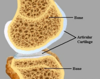
Articular CartilageIn order to understand synovial fluid it is necessary to look at articulated cartilage as both part of the joint are dependent on each other. Articular cartilage is a non mineralised collagen layer which is present where two bones meet in a movable joint. It work with synovial fluid to facilitate low friction movement, damp vibrations and absorb shock.
The articulated cartilage does not contain blood vessels or nerves and relies on the synovial fluid to get nutrients and its oxygen. The tangential layer is porous. When the joint is loaded the pressure squeezes fluid,including waste fluids ,out of the out of the cartilage and when the pressure is relieved the fluid seeps back into the cartilage with oxygen and nutrients. Since cartilage has no nerves it cannot be felt when it is being damaged. Often by the time an injury in cartilage is felt, treatment is too late as cartilage may have be wearing away over a long period.
|
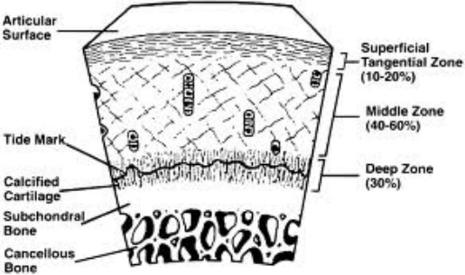
There are three different types of cartilage in the body. The two types seen in the knee joints are hyaline cartilage in the articular cartilage and fibro-cartilage in the meniscus. The hyaline cartilage functions as a low friction, wear resistant tissue designed to bear and distribute loads. The articular cartilage consist of chondrocytes and a dense extracellular matrix composed primarily of water, collagen and proteoglycans. The cartilage in the joint can be split into 4 different layers. The tangential zone has collagen fibers tightly packed parallel to the surface which gives a smooth gliding surface and resists shear. The fibers have a different orientation and size on in the other zone giving more structural properties. The elastic modulas is lowest on the tangential zane and increases inward to the deep zone. This gives a steady increase in deformation from the bone to the contact point which helps with shock absorption and distributing the loads .
|
 |
|
